You have done your mammogram and you were told you have dense breasts. What to Know and What to Do
1. Dense Breast Tissue
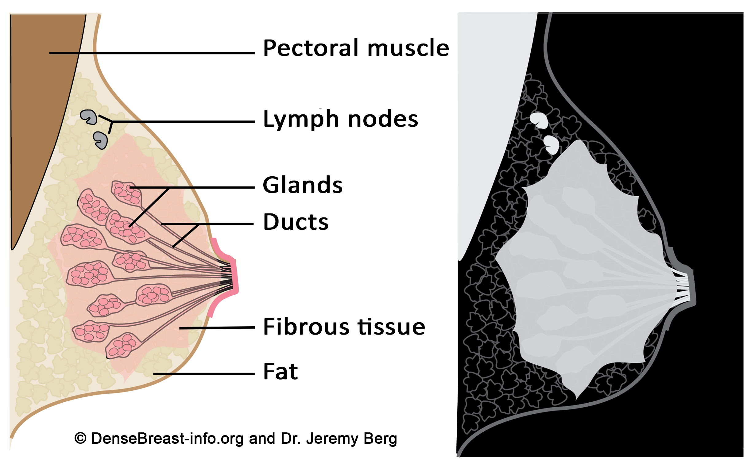
Why it matters: Breasts are made up of a combination of glandular tissue, fibrous (connective) tissue, and fatty tissue. The glandular tissue includes the lobules (which produce milk) and ducts (which carry milk to the nipple). Fibrous tissue provides structure and support, while fatty tissue fills the spaces in between. The proportion of these tissues varies from person to person, and this balance is what determines breast density.
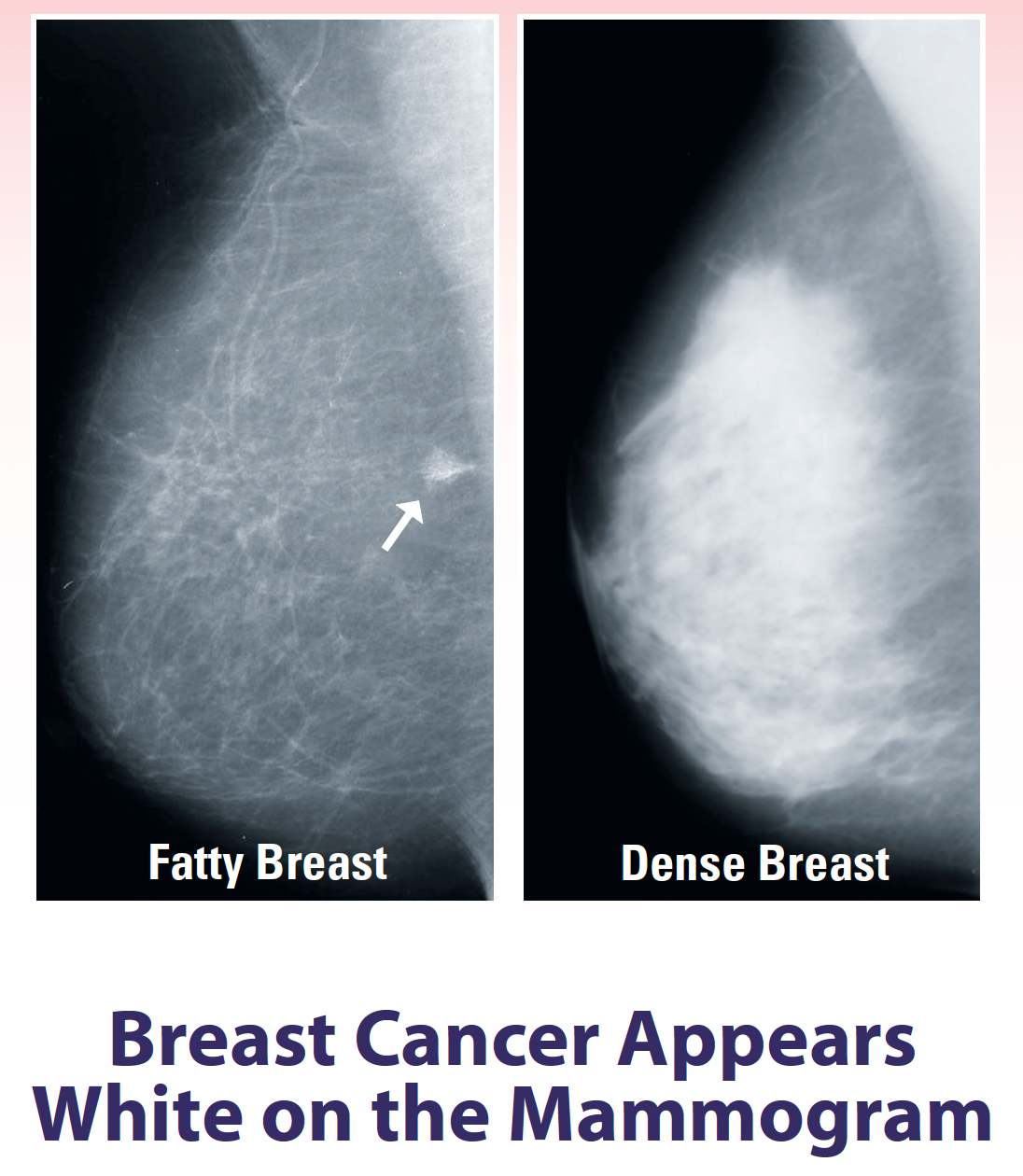
When there is more glandular and fibrous tissue compared to fat, breasts are considered “dense.” Dense breasts are completely normal and quite common—nearly half of women over 40 have them. However, density matters because dense tissue can make it harder for radiologists to see certain changes or small tumors on a mammogram, since both dense tissue and cancer appear white on the image.
Having dense breasts doesn’t mean you have breast cancer, but it does mean you may benefit from additional screening. An ultrasound or MRI can provide clearer imaging in dense tissue, helping detect abnormalities that a mammogram alone might miss. Knowing your breast density allows you and your healthcare team to make more informed choices about the right screening plan for you.
Breast Density
2. Breast Density and Women
Breast density is very common. Research shows that about 40–50% of women over the age of 40 have dense breasts (Mayo Clinic, 2024; American Cancer Society, 2023). This means nearly half of women going in for their routine mammograms may have tissue that makes it harder to detect certain changes.
Dense breasts are perfectly normal and not a disease. They simply mean there is more glandular and fibrous tissue compared to fat in the breast. Because both dense tissue and potential abnormalities appear white on a mammogram, it can be more challenging to spot small tumors or other changes (American Cancer Society, 2023).
For this reason, women with dense breasts may benefit from additional imaging tests such as breast ultrasound or MRI to provide a clearer view (U.S. Food & Drug Administration, 2023). Knowing your breast density helps you and your healthcare team make informed decisions about the best screening plan for you.
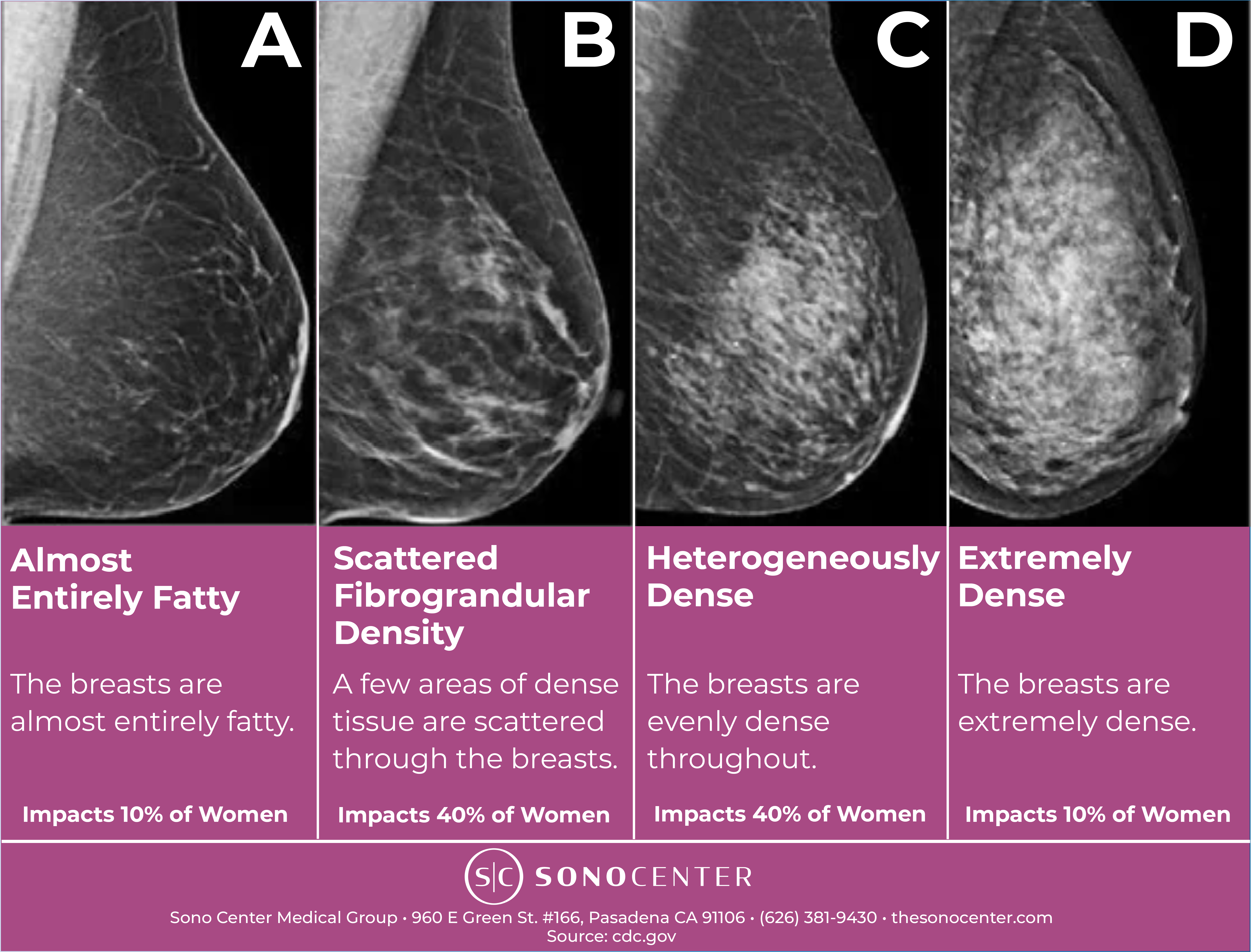
3. Screening and Breast Density
When radiologists look at mammograms, they can see how dense the breasts are. In these four mammogram images, the dense tissue appears as white. Categories A and B are “not dense” breasts. Categories C and D are “dense” breasts.
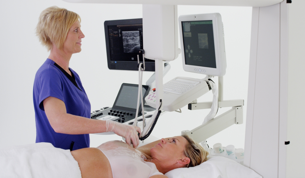
One of the most advanced and effective supplemental imaging options is the SonoCine Automated Whole Breast Ultrasound. Unlike traditional ultrasounds, which are often operator-dependent, SonoCine uses an automated system to capture a complete, consistent view of the entire breast. This technology is particularly beneficial for women with dense breasts, as it allows for better visualization of potential tumors that may be hidden in dense tissue during a standard mammogram.
Dense Breast
4. Dense Breast Tissue Appears White, Similar to Cancer
Why it matters: Dense tissue appears white on a mammogram, and so does cancer. This makes it harder for radiologists to see cancer in dense breasts. Because cancer can be missed, it is often found when larger and at a later stage in women with dense breasts.
Benefits of SonoCine:
Improved Detection Rates: Studies have shown that adding ultrasound to mammography significantly increases the detection of small, invasive cancers that are often missed in dense breasts. The automated nature of SonoCine provides a more comprehensive scan of the entire breast, ensuring nothing is overlooked.
Non-Invasive and Comfortable: The SonoCine ultrasound is a non-invasive, radiation-free exam. It’s painless and comfortable, making it a stress-free experience for women who may be anxious about additional screenings.
Increased Accuracy for Dense Breasts: Dense breast tissue can obscure cancer on a mammogram, but SonoCine’s high-resolution imaging is able to penetrate dense tissue, providing a clearer and more accurate view. This can help catch cancer earlier, when it’s most treatable.
Comprehensive Screening: The automated nature of the SonoCine AWBUS system ensures that the entire breast is scanned thoroughly, reducing the likelihood of human error or oversight. This makes it a more reliable option for detecting abnormalities in dense breast tissue.
5. Risk Factor
Having dense breasts is considered an independent risk factor for breast cancer. Women with very dense breast tissue may be about 1.5 to 2 times more likely to develop breast cancer compared to women with average breast density (American Cancer Society, 2023). Dense tissue not only makes it harder for radiologists to see small changes on a mammogram, but it also may be linked to the way certain cancers develop. While breast density alone doesn’t mean you will get breast cancer, it’s important information that helps guide your personal screening plan. If you have dense breasts, your doctor may recommend additional imaging, like breast ultrasound or MRI, for the most accurate detection.
At Sono Center, we’re not just here to scan—we’re here to support, inform, and empower you. Whether you’re due for a breast screening or looking to understand your broader cancer risk, we’re here for every step of the journey.
Schedule your next screening today and stay proactive about your health.